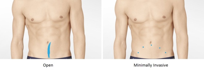
Two cuts are better than one.

2222

Original image by Calvin Klein Inc. – Edited by Richard Kizzee

When told “you will need surgery,” many people feel helpless. If someone is going to look inside your body, you had better have some say in how they do it. Vrije Universiteit Amsterdam is home to a group of researchers who show why opting for minimally invasive gastric surgery may be a safer option than fully open surgery.

Stomach (gastric) cancer is the cause of 10% of cancer deaths worldwide. This type of cancer often calls for total gastrectomy, full removal of the stomach, to be stopped. Two methods generally exist for approaching the stomach to remove it. A single, long incision can be made allowing for surgery through a fist sized hole. Another approach is making multiple small incisions (about wide enough to poke a finger in) for surgery.

Researchers of this study compared open total gastrectomy (OTG) with minimally invasive total gastrectomy (MITG) for markers of surgical quality and patient outcome. This was done by analyzing the results of 12 studies involving a total of 1,360 patients, of whom 768 underwent OTG and 592 MITG. The results of their meta-study showed (average differences in parentheses):

Shorter operation time: OTG (48.06 minutes faster)
Lower blood loss: MITG (160.70 mL less)
Lower time to recovery: MITG (1.05 days sooner)
Shorter length of hospital stay: MITG (2.43 days less)
Less postoperative complications: MITG (32.72% less)
Completeness of procedure: Equal

Both techniques were equally excellent at removing cancer from the patients, however, the minimally invasive technique left people recovering faster and with less complications after surgery. The benefits of MITG seen here may also apply to da Vinci robotic surgery in general.

If you or a loved one need surgery, remember that techniques are improving every day. Whether opting for open or minimally invasive (or even robotic) surgery, you are in good hands.

Reference:
Jennifer Straatman, Nicole van der Wielen, Miguel A. Cuesta, Elly S. M. de Lange – de Klerk, Elise P. Jansma, Donald L. van der Peet. Minimally Invasive Versus Open Total Gastrectomy for Gastric Cancer: A Systematic Review and Meta-analysis of Short-Term Outcomes and Completeness of Resection. World Journal of Surgery, 2016.
DOI: 10.1007/s00268-015-3223-1

I’m not bipolar, my mother had my blood tested.

1212

Carrie Mathison – Monique Nazareth

Assessing mood is inherently subjective.
“I’m sad.”
– “How sad are you?”
“Very sad.”
– “. . .”
However, scientifically quantifiable means of determining the severity of mood disorders are on the rise. Through the cooperation of researchers of the United States, Europe, and Australia spanning from the University of Texas to Deakin University, a blood compound (BDNF) was identified as an indicator of bipolar mood swings.

Brain-derived neurotrophic factor (BDNF) is a protein that aids in the development and survival of neurons. BDNF works to promote growth of neurons and synapses in the brain, eyes, and kidneys among other places. This protein has important links to obesity, epilepsy, schizophrenia, and as seen in this study, bipolar disorder.

Bipolar disorder causes intense emotionally states (overly happy or extreme hopelessness) that last for unusually long periods of time. In a state of mania, one might be easily distracted, sleep very little, overestimate their personal abilities, and act impulsively on high risk high pleasure activities. During a depressive episode the person might feel tired, lack concentration, drastically change eating habits, and actively think of death or suicide.

The researchers of this study analyzed the data of 52 studies published from 2005 to 2015 that looked at levels BDNF circulating within 3,339 individuals in manic, depressive, or euthymic (moderately happy) states compared to 3,142 healthy control participants. The goal was to find a link between amount of BDNF in the blood and current manic or depressed signs. The meta-analysis showed BDNF levels significantly dropped during manic and depressed episodes (Hedges’ g = -0.57 and -0.93 respectively), while euthymia showed no change in BDNF.

Not only did BDNF decrease more with increasing severity of bipolar episode, but individuals treated for their manic episodes showed increases in BDNF with medication. The BDNF increase was not seen with depressive episode treatment, but this could be due to depression signs persisting through treatment (Read as: science is good at depressing people, but bad at cheering them up).

If further research finds that BDNF levels are decreasing before depressive or manic episodes, this protein could be used as a forecaster of bipolar mood jumps. BDNF currently shows the most promise as a biomarker for activity.

In conjunction with other blood assays, BDNF may be able to help determine if in fact someone is bipolar, how severe their mood swings are, and give a more numeric answer to “how sad are you?”.

Reference:
Brisa S. Fernandes, Marc L. Molendijk, Cristiano A. Köhler, Jair C. Soares, Cláudio Manuel G. S. Leite, Rodrigo Machado-Vieira, Thamara L. Ribeiro, Jéssica C. Silva, Paulo M. G. Sales, João Quevedo, Viola Oertel-Knöchel, Eduard Vieta, Ana González-Pinto, Michael Berkand, André F. Carvalho. Peripheral brain-derived neurotrophic factor (BDNF) as a biomarker in bipolar disorder: a meta-analysis of 52 studies. BioMed Central Medicine, 2015.
DOI: 10.1186/s12916-015-0529-7

Love and Biology

From Ihle, Kempenaers, and Forstmeier

From Ihle, Kempenaers, and Forstmeier

If two people marry each other to mutually benefit each other’s rise to the top of the political ladder or because their combined wealth will give their child a worry free life, what’s love got to do with it? If a man married his wife because his mother arranged it, would that affect the affection he showed towards their son? Researchers of the German Max Planck Institute for Ornithology studied zebra finches to find out if being able to freely choose one’s mate, ensured reproductive success.

The zebra finch is a monogamous species of bird that tends to stay with its chosen mate for life. With only one mate, however, the rate at which baby finches can be produced is limited. With the goal of creating offspring that will continue to pass down genes, many organisms including lions, deer, and crickets choose the polyandrous route and mate with numerous partners. The married zebra finch is not at a disadvantage, however. Fitness is a means of gauging success in passing on genes. This study found the relative fitness of finch pairs that chose their mate and compared it to those whose mate was chosen for them.

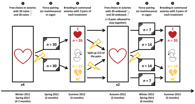
160 zebra finches freely chose a partner of the opposite sex. Following, half of the pairs were assigned to a random partner instead of their chosen one. Two more rounds of either allowing pairs to stay together or force choosing a partner for them followed this. After each pairing cycle, the couples were allowed to court and mate (get to know each other and have sex). Either good parenting or good genes would be the factor to determine if the group of finches choosing their own mate or the group with their mate chosen for them as having more fitness.

The study found the pairs of finches whom chose their own partners (C) were 37% more reproductively fit than pairs with mates force chosen for them (F). Many factors played into this:

– (C) vs. (F)
– Divorce rate: 8% vs. 24%
– Disappeared or buried eggs: 12% vs. 19%
– Offspring mortality: 32% vs. 52%
– Male courting outside his pair: Once every five hours vs. once every three hours

The higher fitness of zebra finch pairs that chose their own mates (C) supports the behavioral compatibility model. The finch pairs may have chosen each other based on harmony in parenting or general behavior according to the study. The downfalls seen within the (F) pairs were lower nest attendance by males, reduced motivation to care for young, chronic stress with a forced mate, lower female responsiveness, and higher male cheating rates with compatibility not increasing as marriages lengthened.

Is love the ultimate motivator for ensuring family success? Studies with people follow similar conclusions to this study in that emotionally satisfying relationships showed more commitment and less domestic violence. Lions can have their harem. I for one am a proponent of love (and so is science it seems).

Reference:
Malika Ihle, Bart Kempenaers, Wolfgang Forstmeier. Fitness Benefits of Mate Choice for Compatibility in a Socially Monogamous Species. PLOS Biology, 2015.
DOI: 10.1371/journal.pbio.1002248

Resveratrol Vs. Exercise

Photo provided by  Anthony L. Hall

Photo provided by Anthony L. Hall

Exercise proves itself yet again as a more powerful way to stay healthy than taking dietary supplements. The department of sport science at Chungnam National University is where research was done to study the effectiveness of exercise versus resveratrol on fighting fat.

Resveratrol is a nutrient found in many foods that has become famous in recent years for its benefits of reducing blood pressure, protecting the heart, and preventing neuro-degenerative diseases. Specifically related to this research, resveratrol can increase metabolism, reduce lipids in the body, and help weight loss. It has been touted as an alternative to exercise.

To test this, 30 obese mice were assorted into three groups of 10: (R) mice were given resveratrol five days per week (10mg/kg of body weight), (E) mice exercised (30-60 minutes of treadmill running for five days a week), (C) mice acted as the control. All mice were on a low fat diet for eight weeks along with their group’s regimen during the study. The concluding results showed an average weight loss of 15% for (E) mice and about 5% for (R) compared to the controls (C). The higher weight loss most likely resulted from increased fat breakdown and blockage of new fat creation seen from exercise.

Although resveratrol has many highly sought after health benefits, it cannot beat good ol’ fashioned exercise in the battle against obesity.

Reference:
Jun Hyun Jeong, Hee Geun Park, Young Ran Lee, Wang Lok Lee. Moderate exercise training is more effective than resveratrol supplementation for ameliorating lipid metabolic complication in skeletal muscle of high fat diet-induced obese mice. The Journal of Exercise Nutricion & Biochemistry, 2015
http://dx.doi.org/10.5717/jenb.2015.19.2.131

Arm fractures followed soon by death

Photo provided by Andre van Schalkwyk of Antilogic

Photo provided by Andre van Schalkwyk of Antilogic

Broken arm? No problem! One trip to the hospital, and you’re ready to continue your long healthy life. Maybe not. University of Helsikinki’s very own have reported a strong link between upper limb fracture and increased rate of death.

Just shy of 6,000 people of Finland who sustained bone fractures to their upper extremities were followed for an average of six years between 2002 and 2012. Researchers wanted to find out if those with shoulder, arm, wrist, etc. fractures had an increased risk of death similar to those with hip fractures.

Previous studies have documented that people with one or more hip fractures have nearly a three times higher rate of dying than people of the same age without a hip fracture. The standardized mortality ratio (SMR) was 2.18 for women and 3.17 for men. The SMR is the amount of deaths of a studied population as compared to the general population. An SMR of 1 would mean an equal rate of death in both populations, while an SMR of 2 would mean 100% more cases of death in the studied population.

This study found that 12% of the studied population had died five years after their upper extremity fracture. For individuals between 16 and 59 years old, there were nearly 200% more cases of death for individuals with a bone fracture as compared to those without (SMR: 2.2 for women, 3.0 for men). Individuals 60 years of age and older had an average of 40% more cases of death (SMR: 1.2 for women, 1.6 for men). If the fracture was in the individual’s humerus, the arm bone that extends from shoulder to elbow, mortality was much higher for both sexes of all ages.

Leading causes of death for individuals sustaining upper limb fractures were cardiovascular disease, accidents, and violence. No, this does not mean a broken arm increases risk of heart attack (except in the mother of the son who broke his arm playing street hockey), but concern should be raised after sustaining fracture.

People who break an arm, or sustain similar upper extremity fractures, may be more frail than others, leading a risky lifestyle, or just unlucky. Whatever the reason, this study says be careful in the future and stay healthy!

Reference:
Axel Somersaloa, Juha Palonevab, Hannu Kautiainencd, Eija Lönnroose, Mikko Heinänenf, Ilkka Kivirantaf. Increased mortality after upper extremity fracture requiring inpatient care. Acta Orthopaedica, 2015.
http://dx.doi.org/10.3109/17453674.2015.1043833

Herpes don’t hurt me!

Courtesy of Emneth Design

Courtesy of Emneth Design

“Don’t look at me, I’m a monster!” The herpes simplex virus (HSV) plagues many people worldwide putting them at social and more importantly physical danger as the virus can lead to mortal complications. Researchers of the University of Washington and Hutchinson Cancer Research Center found that Nelfavir can trap herpes within cells and turn it from a danger to an annoyance.

This study looked into the ways in which Nelfinavir (NFV), an anti-cancer drug, could be used to combat the virus causing herpes, specifically HSV-1. HSV is a non-living, infectious agent that looks like a durian fruit. The DNA of the virus is inside while the outside “skin” is made of protein and “spikes” are made of glycoproteins (sugar-proteins). The glycoproteins are what allow the virus to enter human cells and infect. HSV infection causes the cell to waste its resources to make new viruses that can infect other cells. With HSV comes elevated risk of aquiring HIV and Kaposi sarcoma, a cancer.

Imagine a factory that makes cars and all the machinery inside is blue. The factory can be equated to a cell. If a virus infects, this would be akin to someone adding instructions and an extra machine or two to the factory that now allows bombs to be made in the car factory. Imagine the extra machine is yellow. It is easy to spot and get rid of the extra machinery that is making bombs if it does not look like the original factory machinery. This is how most drugs target and destroy viruses. The virus machinery is often easy to differentiate from cell machinery. What if the virus machinery were also blue? This is when a virus becomes drug resistant. The virus has changed, so the drug can no longer target and destroy it. Instead of targeting the virus itself, another method of stopping viruses is by changing the cell machinery so that it will rarely make a fully functioning bomb (virus). This is how NFV acts on HSV-1.

The researchers infected human connective tissue cells, fibroblasts, with HSV-1 and treated with NFV to measure its effectiveness in blocking virus production in vitro. It was found that NFV blocked viral protein maturation, glycoprotein development, and viral packaging.

Virus retention inside the cell was 79:14, NFV treated:untreated
Virus particles found outside the cell was 3:51, NFV treated:untreated
Virus completion inside the cell was 0:9, NFV treated:untreated

This meant that although HSV-1 was getting into the cells, new viruses were created broken and could not leave or infect other cells. It was also found that HSV-1 was not becoming resistant to the drug even after subjected to drug pressure. This is because NFV affects the cell rather than virus.

This newfound use for Nelfinavir shows promise for longterm control of herpes. This ubiquitous and malignant virus may soon be just a small bother.

Reference:
Soren Gantt, Eliora Gachelet, Jacquelyn Carlsson, Serge Barcy, Corey Casper, Michael Lagunoff. Nelfinavir Impairs Glycosylation of Herpes Simplex Virus 1 Envelope Proteins and Blocks Virus Maturation. Advances in Virology, 2015
http://dx.doi.org/10.1155/2015/687162

Stem Cells Heal Hearts

Photo courtesy of Abigail Droege

Photo courtesy of Abigail Droege

Running up stairs, hiking a new trail, eating great food can all cause acute, unexpected heart attack. That is, given arterial plaque buildup is unstable and ready to pop. Researchers of Zhejiang University, Ningbo University, and the Chinese PLA 305 Hospital, however, found that mesenchymal stem cells can stabilize this heartbreaking plaque.

Cardiovascular disease is the number one most common cause of death worldwide accounting for over 25% of all deaths in 2012. More specifically, one out of every six deaths in the United States is caused by heart attack alone. A large contributing factor to heart attack is accumulated plaque within the walls of arteries. This research focused on a big player in sudden and often unexpected heart attack, vulnerable plaque.

As cholesterol moves through the bloodstream, excess will often accumulate in the walls of blood vessels. Seen as a foreign substance, the cholesterol pool will constantly be attacked by white blood cells. This however, leads to larger plaque buildup as the macrophages and neutrophils are swallowed up by the tarlike plaque. As well, the fibrous cap keeping the plaque away from the blood thins out as cells die. Although the narrowing of arteries is dangerous, acute heart attack is only induced once the lipid deposit ruptures and spills into the bloodstream. This instantly clogs arteries.

Process of vulnerable plaque rupture and arterial blockage

Process of vulnerable plaque rupture and arterial blockage

If inflammation around the plaque could be stopped, white blood cells would not exacerbate growth, the fibrous cap would remain thick and robust, and the fatty clot would not form. The researchers experimented with stem cells’ ability to prohibit inflammation and thereby stabilize plaque deposits.

Forming groups of ten, 30 rabbits were put into either stable plaque (SP), vulnerable plaque (VP), or mesenchymal stem cell (MSC) groups. The groups had plaque formations with no danger of rupture, extreme danger of rupture, or extreme danger of rupture with the addition of MSC’s respectively. To detect plaque stability throughout the project, H&E and Masson staining were used to measure the fibrous cap/lipid core ratio. A high ratio indicated low chance of rupture (stable) while a low ratio indicated high chance of rupture (vulnerable).

After the intravenous introduction of MSC’s, levels of various inflammation markers were measured over a four week period. The following was found:

Pro-inflammatory compound levels (hs-CRP, TNF-a, IL-6, NF-kb)
MSC: Low
SP: Low
VP: High

Anti-inflammatory compound levels (TSG-6, IL-10)
MSC: High
SP: Normal
VP: Normal

As well, the levels of cellular suicide inducer MMP were found to be low in the MSC group. This meant that the stem cells were decreasing inflammation and apoptosis at the site of the plaque deposit. Vulnerable plaques with high risk of inducing abrupt heart attack were transformed into less harmful, stable plaques without the risk of causing sudden cardio-ischemic attacks.

Stem cells seem to be the next big hero of the heart. The list of accomplishments for these little multipotent warriors just keeps on growing.

Reference:
Shuang-shuang Wang, Si-wang Hu, Qing-hua Zhang, Ai-xiang Xia, Zhi-xin Jiang, Xiao-min Chen. Mesenchymal Stem Cells Stabilize Atherosclerotic Vulnerable Plaque by Anti-Inflammatory Properties. PLOS ONE, 2015
http://dx.doi.org/10.1371/journal.pone.0136026

Humidity > Influenza Virus

Photo, James Gathany Photo credits: CDC

Photo, James Gathany
Photo credits: CDC

Each year in the winter influenza or “flu” season rolls around and this sickness runs rampant throughout the country. This virus, and many in general, are quite scary because of how easily they can be spread through the air ie. via coughing. Researchers of the National Institute for Occupational Safety and Health (NIOSH of the CDC) have recently linked higher humidity and transmission particle size to more inactivation of influenza (and less spread).

Influenza is an RNA virus that affects mammals and birds causing a small scale pandemic every year in the United States. Viruses tend to mutate very quickly, therefore, it is hard to find a single band-aid to blanket cure this bug. Most people can spread the virus between -1 and 6 days after showing symptoms. Airborne transmission is most likely when this virus appears since many people are in closed quarters attempting to stay warm at home with others during winter.

To test the rate at which influenza stays infectious in different humidities, researchers set up coughing and breathing test dummies in an isolated room. The sneezing dummy released about 4.5×10³ virus/liter of air into the room, where 4.6% of that virus was seen to be infectious upon discharge. Of that, upwards of 77.2% of the virus retained infectivity at lower relative humidity (7-23%), while only 14.6% retained infectivity at higher relative humidity of 43%. As a grounding point, average in home humidity during winter is just above 20%, while summer humidity indoors is just below 60%.

Next, the rate of loss of infectivity was measured comparing low and moderate humidity (20% and 45%). Within 15 minutes, 52% of the virus’ infective ability was lost at 45% humidity compared to 20% humidity.

Together these two showed more virus lost its ability to infect new people/etc. at higher relative humidities.

Finally, transmission particle size was measured to see if it had bearing on influenza’s spread and infection at differing humidities. Three particle sizes were used, <1 µm, 1-4 µm, and >4 µm. At the two extremes, <1 & >4 µm, 94% infectivity was seen as lost in the first 15 minutes after coughing virus into the air. However, the 1-4 µm range of particles only showed 29% decrease in infectivity of the virus after 15 minutes. All sizes did, however, show increased loss of infectious ability with more time passed.

It seems as though humidity has great bearing on the inactivation/spreading ability of the influenza virus. At higher humidities, 45% +, the influenza virus showed very little strength in infecting new hosts. This was doubly true when the particle size they rode on was really small or really big.

The best way to minimize your chance to get influenza each year without getting a flu shot?…move to a state with really high humidity in the winter.

Reference:
John D. Noti, Francoise M. Blachere, Cynthia M. McMillen, William G. Lindsley, Michael L. Kashon, Denzil R. Slaughter, Donald H. Beezhold. High Humidity Leads to Loss of Infectious Influenza Virus from Simulated Coughs. PLoS ONE, 2013.
http://dx.doi.org/10.1371/journal.pone.0057485

unOC linked to Diabetes relief

GLP-1 effects on different human organs.
Photo credits: Daniel J. Drucker (author of the paper of
DOI: 10.1016/j.cmet.2006.01.004)

Researchers have perhaps found another means by which to manage type 2 diabetes. A group from 九州大学 (Kyushu  University) has shown that addition of uncarboxylated osteocalcin (unOC) stimulates glucagon-like peptide-1 (GLP-1) release and insulin secretion in mice.

GLP-1 is normally released in released into the blood in response to eating, so that insulin can be released and digested glucose can be taken into cells. In those with type-II diabetes, the insulin release pathway is impaired in some way, so insufficient insulin (or no insulin) is produced to promote glucose uptake.

UnOC has shown in previous studies to have an effect on the pancreas directly to stimulate insulin release; therefore, this study wanted to test the indirect effect of unOC on insulin secretion via the GLP-1 pathway.

Using mice, unOC’s effect on GLP-1 secretion was tested. 20 minutes after giving mice unOC orally or intraperitoneally, a maximal rise of 50 pg/mL of serum GLP-1 was seen. However, this significant rise was only seen in mice also given the DPP-IV inhibitor sitgaliptin. This needed to be co-administered because GLP-1 has a very short half life and would breakdown before the effects could be seen.

Extending from this, the insulin levels in mice given unOC were seen as raised as well. With the combination of unOC and sitagliptin, insulin rose (maximally) 0.4 ng/mL. These results were seen administering therapies orally or via intrapineal injection. Administering intravenously gave similar results with significant rises in GLP-1 and insulin sooner (as expected).

This study shows unOC may be a strong therapy for type II diabetes management if given to patients in the pure chemical form, as an additive to food, or the like.

Reference:
Akiko Mizokami, Yu Yasutake, Jing Gao, Miho Matsuda, Ichiro Takahashi, Hiroshi Takeuchi, Masato Hirata. Osteocalcin Induces Release of Glucagon-Like Peptide-1 and Thereby Stimulates Insulin Secretion in Mice. PLoS ONE, 2013.
http://dx.doi.org/10.1371/journal.pone.0057375

————————-

A note from me:

Lately my writings have been sluggishly drawn out and without the wherewithal to inspire (not to mention I’ve been posting suuuuuper late). Because of this, I’m taking a week break from blogging. For those that follow: I’ll see you on March 1st. For those that stumbled upon this today: Hopefully you’ll muse my articles, learn some cool stuff, and come back in a week when I pick up again.

See you all seven mornings from now 🙂

Retinal degeneration and blindness, possibly preventable

Photo by Aaron (~Sayn)

Neurodegenerative diseases such as retinitis pigmentosa (RP) currently ravage through the eyes of millions of people worldwide unhindered and untreated. Genetic disorders like these often leave patients blind with no good news from the time of diagnosis. That is likely to change soon as researchers of the Universities of Toronto, Lausanne, Lund, and The Centre of Biomedical Genetics (Amsterdam) found Bmi1 and cyclin-dependent kinases (CDKs) a potential targets to prevent blindness caused by neurodegeneration.

Retinal pigmentosa results from the mutation of around 200 loci or genes leading to the loss of rod and cone photoreceptors. Previous studies showed that neurons prepared to die (due to a neurodegenerative disease) reexpress cell cycle-related proteins like CDKs. Specifically CDK4 and CDK6 activation was seen as involved with neuronal death in these conditions.

Bmi1 to E2F1 cycle

Bmi1 is a polycomb group protein (proteins that silence genes) which inhibits CDK inhibitors. These normally lessen the activity of CDK’s, which inhibit retinoblastoma (Rb) proteins. The Rb proteins down-regulate E2F1, which causes the cell to divide past it’s prime. This little cell cycle here has been shown to be the cause of neurodegeneration in Parkinson’s disease; therefore, researchers are investigating the effects of it in ophthalmological cases.

Rb1 mice (those with induced RP) were first analyzed for their expression of cell cycle markers. Between postnatal day nine (P9) and P12, the amount of CDK4/6 significantly increased in the apoptotic cells of the Rd1 mice. However, almost no CDK4/6 was detected in the wild type mice’s retinal cells.

Because of the higher expression seen, CDK interference was run. An ex vivo (pulled part of the retina out of the mouse to study) assay was performed with retinal explants of Rd1 mice. Using roscovitine (CDK inhibitor) vs. no treatment groups, photoreceptor degeneration was measured. The roscovitine group showed 42% less apoptotic photoreceptors. This meant CDK4/6 were contributing to the loss of photoreceptors.

Consistent with this, inhibited Rb1 was found in Rb1 mice and not wild type mice. This meant E2F proteins were allowed to freely push the cell into apoptotic division. Playing with alleles and expression patterns of E2F1,2,3 in combination, researchers were able to determine ESF1 was more important than E2F2/3 in neurodegeneration. Deleting the gene resulted in slowed, but not stopped photoreceptor degeneration.

Finally, Rd1/Bmi1-/- mice were analyzed vs. Rd1 mice to see the protective effect of removing Bmi1 all together. Respectively, 40% and 90% photoreceptor loss was seen in the mice. Rd1/Bmi1-/- mice lost 50% less photoreceptors than the normal retinitis pigmentosa mice.

Pulling all together, retinal degeneration was significantly slowed and prevented leading to a protection of photoreceptors by inhibition of Bmi1, CDK4/6, and E2F1. Being diagnosed with RP may not be as hopeless any longer. Once therapies are pushed into the clinic using the targets found in this study, RP patients may shrug at their diagnosis rather than shudder.

Reference:
Dusan Zencak, Karine Schouwey, Danian Chen, Per Ekström, Ellen Tanger, Rod Bremner, Maarten van Lohuizen, Yvan Arsenijevic. Retinal degeneration depends on Bmi1 function and reactivation of cell cycle proteins. Proceedings of the National Academy of Sciences of the United States of America, 2013.
http://dx.doi.org/10.1073/pnas.1108297110